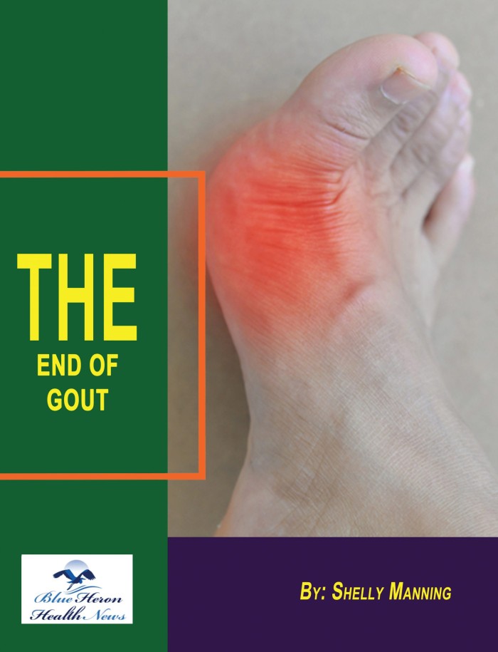
How Gout is Diagnosed in a Clinical Setting
Diagnosing gout in a clinical setting involves a combination of patient history, physical examination, laboratory tests, and imaging studies. Here’s an overview of the key steps and methods used in the diagnosis of gout:
1. Patient History
- Symptoms: The clinician will ask about the patient’s symptoms, focusing on joint pain, swelling, and redness. Gout typically presents with sudden, severe pain, often starting at night, particularly affecting the big toe (podagra).
- Previous Episodes: Inquire about the frequency and duration of gout attacks, as well as any previous diagnoses or treatments.
- Medical History: Review the patient’s medical history for conditions associated with gout, such as kidney disease, hypertension, diabetes, and metabolic syndrome.
- Lifestyle Factors: Discuss dietary habits, alcohol consumption, and medication use (e.g., diuretics) that may contribute to hyperuricemia and gout.
2. Physical Examination
- Joint Assessment: The clinician will examine affected joints for signs of inflammation, including redness, swelling, warmth, and tenderness. The first metatarsophalangeal joint is commonly affected but gout can also occur in other joints.
- Tophi: Look for the presence of tophi, which are deposits of uric acid crystals that can develop in chronic cases and are often found in the ears, fingers, and around joints.
3. Laboratory Tests
- Serum Uric Acid Levels: A blood test to measure uric acid levels is commonly performed. While hyperuricemia (elevated uric acid) can indicate a risk for gout, it is not definitive since some individuals with gout may have normal uric acid levels during an acute attack.
- Joint Aspiration (Arthrocentesis): The most definitive test for diagnosing gout is the aspiration of synovial fluid from an inflamed joint. The fluid is then analyzed for the presence of monosodium urate crystals using polarized light microscopy.
- Crystal Analysis: The identification of needle-shaped, negatively birefringent crystals confirms the diagnosis of gout.
- Complete Blood Count (CBC): A CBC may be performed to check for signs of infection or other underlying conditions.
4. Imaging Studies
- X-rays: While not used for diagnosis, X-rays may be performed to rule out other joint conditions or assess joint damage in chronic gout.
- Ultrasound: Musculoskeletal ultrasound can help visualize urate crystals in the joint or soft tissues and may be useful in ambiguous cases.
- Dual-Energy Computed Tomography (DECT): This advanced imaging technique can identify uric acid deposits in joints and soft tissues, providing additional support for the diagnosis.
5. Differential Diagnosis
- Rule Out Other Conditions: The clinician will consider and rule out other conditions that may cause similar symptoms, such as pseudogout (calcium pyrophosphate crystal deposition disease), septic arthritis, osteoarthritis, and other types of inflammatory arthritis.
6. Diagnosis Confirmation
- Clinical Criteria: The American College of Rheumatology (ACR) criteria for the diagnosis of gout include a combination of clinical, laboratory, and imaging findings. A confirmed diagnosis typically relies on the presence of urate crystals in the synovial fluid or a history of recurrent gout attacks along with hyperuricemia.
Conclusion
A comprehensive approach, combining patient history, physical examination, laboratory tests, and imaging, is essential for accurately diagnosing gout in a clinical setting. Early diagnosis and appropriate management are crucial to relieve symptoms, prevent future attacks, and avoid potential complications associated with chronic gout.
The Bone Density Solution by Shelly ManningThe program is all about healthy food and healthy habits. As we discussed earlier, we develop osteoporosis due to low bone density. Therefore, you will have to choose the right food to help your calcium and other vitamin deficiencies. In addition to healthy food, you will have to regularly practice some mild exercises. Your doctor might offer you the same suggestion. However, the difference is that The Bone Density Solution will help you with an in-depth guide.

