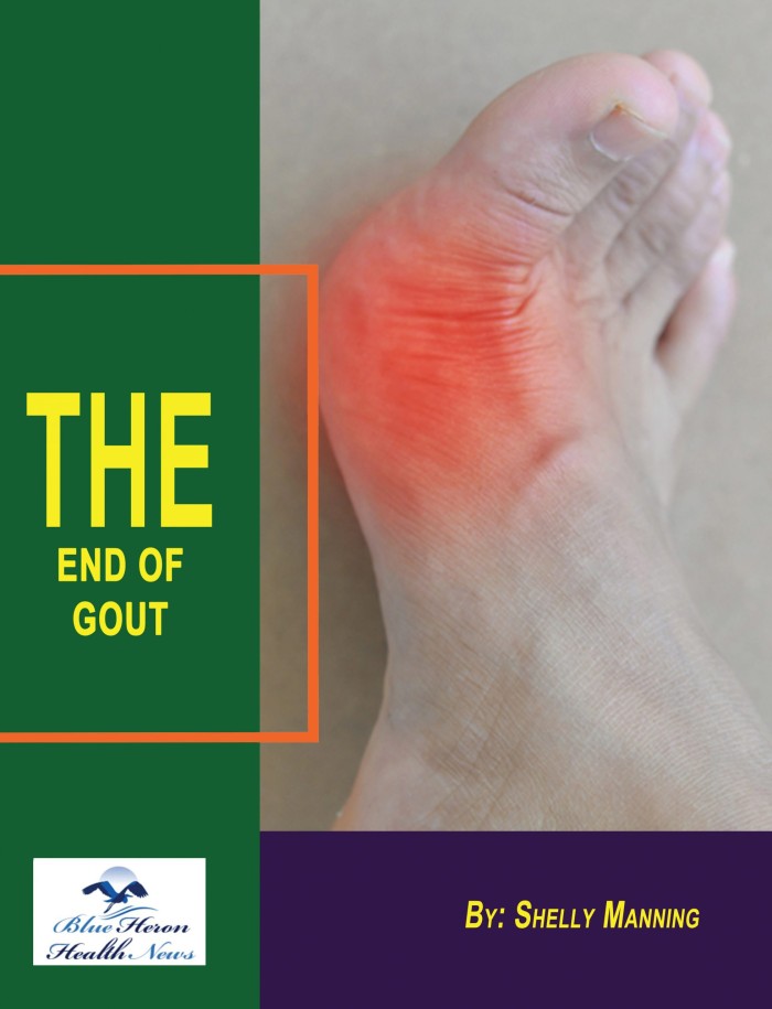
The End Of GOUT Program™ By Shelly Manning Gout has a close relation with diet as it contributes and can worsen its symptoms. So, it is a primary factor which can eliminate gout. The program, End of Gout, provides a diet set up to handle your gout. It is a therapy regimen for gout sufferers. It incorporates the most efficient techniques and approaches to be implemented in your daily life to heal and control gout through the source.
How Gout is Diagnosed
Diagnosing gout involves a combination of clinical evaluation, laboratory tests, and imaging studies to confirm the presence of the disease and distinguish it from other types of arthritis. Here’s an overview of how gout is diagnosed:
1. Clinical Evaluation
- Medical History: The doctor will start by taking a detailed medical history, asking about the patient’s symptoms, frequency of attacks, and any relevant personal or family history of gout or other conditions like kidney disease, hypertension, or obesity.
- Symptom Assessment: The hallmark symptom of gout is a sudden, severe attack of pain, redness, warmth, and swelling in a joint, most often the big toe. The doctor will inquire about the onset, duration, and severity of the symptoms, as well as any triggers (such as diet or alcohol consumption).
- Physical Examination: The affected joint(s) will be examined for signs of inflammation, such as swelling, redness, tenderness, and limited range of motion. If the patient has tophi (hard nodules formed by uric acid crystals), these may be visible under the skin around joints or in other areas such as the earlobes.
2. Laboratory Tests
- Serum Uric Acid Test: A blood test is conducted to measure the level of uric acid in the blood. While elevated uric acid levels (hyperuricemia) can indicate gout, it’s important to note that not everyone with high uric acid levels has gout, and some people with normal levels may still have gout. Therefore, this test alone is not definitive but provides useful information.
- Synovial Fluid Analysis: The most definitive test for diagnosing gout involves extracting fluid from the affected joint (a procedure known as arthrocentesis) and examining it under a microscope. The presence of monosodium urate crystals in the synovial fluid is a clear indicator of gout. This test helps differentiate gout from other types of arthritis, such as rheumatoid arthritis or septic arthritis.
- Blood Tests: Additional blood tests might be conducted to rule out other conditions that can cause joint inflammation, such as rheumatoid arthritis (testing for rheumatoid factor and anti-CCP antibodies) or infection (checking white blood cell counts).
3. Imaging Studies
- X-Rays: X-rays are typically not necessary for diagnosing early gout but may be used in chronic or advanced cases to assess joint damage. X-rays can reveal characteristic changes in the bone, such as erosions or tophi, particularly in long-standing gout.
- Ultrasound: Ultrasound is increasingly used to detect urate crystals in the joints and soft tissues. It can visualize the “double contour sign,” which indicates the presence of urate crystals on the surface of cartilage. Ultrasound is non-invasive and can help in diagnosing gout without needing to extract joint fluid.
- Dual-Energy CT (DECT): DECT is a more advanced imaging technique that can specifically identify urate crystals in the joints and soft tissues. It provides a detailed image and is particularly useful in complex cases where the diagnosis is uncertain or when tophi are suspected in atypical locations.
4. Differential Diagnosis
- Exclusion of Other Conditions: Gout can mimic other forms of arthritis, so the doctor will rule out conditions such as:
- Pseudogout: Similar to gout but caused by calcium pyrophosphate dihydrate (CPPD) crystals rather than uric acid crystals.
- Septic Arthritis: An infection in the joint that requires immediate treatment.
- Rheumatoid Arthritis: An autoimmune condition that causes chronic inflammation in multiple joints.
- Osteoarthritis: A degenerative joint disease that might present with joint pain and stiffness but lacks the acute inflammatory episodes seen in gout.
5. Clinical Guidelines
- American College of Rheumatology (ACR) Criteria: The ACR provides guidelines for diagnosing gout, which include a combination of clinical symptoms, laboratory findings, and imaging results. The presence of monosodium urate crystals in joint fluid remains the gold standard for diagnosis.
- European League Against Rheumatism (EULAR) Recommendations: EULAR also offers recommendations for diagnosing gout, emphasizing the importance of crystal identification, clinical symptoms, and imaging studies in establishing a diagnosis.
Summary
Diagnosing gout requires a careful combination of clinical assessment, laboratory tests, and imaging studies. While elevated uric acid levels and the presence of monosodium urate crystals in joint fluid are key indicators, it’s essential to distinguish gout from other conditions with similar symptoms. Early and accurate diagnosis is crucial for effective treatment and prevention of complications associated with gout.
The End Of GOUT Program™ By Shelly Manning Gout has a close relation with diet as it contributes and can worsen its symptoms. So, it is a primary factor which can eliminate gout. The program, End of Gout, provides a diet set up to handle your gout. It is a therapy regimen for gout sufferers. It incorporates the most efficient techniques and approaches to be implemented in your daily life to heal and control gout through the source.

