What are the symptoms of gout?
Gout is characterized by various symptoms that can vary in intensity and duration. Here are the primary symptoms associated with gout:
1. Intense Joint Pain
- Common Sites: Gout typically affects the large joint of the big toe, but it can also occur in other joints such as the ankles, knees, elbows, wrists, and fingers.
- Onset: The pain usually begins suddenly, often at night, and can be severe.
- Duration: Initial attacks generally last between 3 to 10 days. Subsequent attacks might last longer and affect more joints.
- Sources: Mayo Clinic – Gout Symptoms, Arthritis Foundation – What Is Gout?
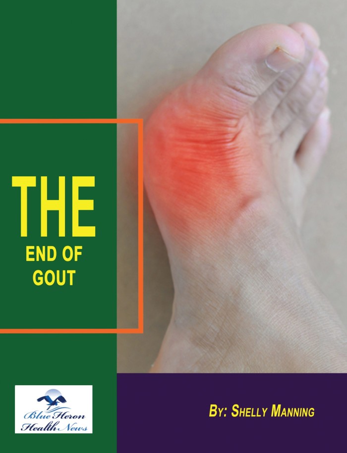
2. Inflammation and Redness
- Symptoms: The affected joint or joints become swollen, warm, tender, and red. This inflammation is due to the body’s immune response to the urate crystals in the joint.
- Sources: National Institute of Arthritis and Musculoskeletal and Skin Diseases (NIAMS) – Gout, Cleveland Clinic – Gout
3. Lingering Discomfort
- Post-Attack Symptoms: After the most severe pain subsides, some joint discomfort may last from a few days to a few weeks. Later attacks are likely to last longer and affect more joints.
- Sources: Mayo Clinic – Gout Symptoms
4. Limited Range of Motion
- Impact on Joints: As gout progresses, it can cause a decrease in the range of motion in the affected joints, making it difficult to move them normally.
- Sources: Arthritis Foundation – Managing Gout
5. Tophi Formation
- Description: With recurrent gout attacks, deposits of urate crystals, called tophi, can form under the skin in nodules. These tophi can occur in various areas such as the fingers, hands, feet, elbows, or Achilles tendons.
- Impact: Tophi are usually not painful but can become inflamed and tender during gout attacks.
- Sources: Mayo Clinic – Gout Symptoms, Cleveland Clinic – Gout
Conclusion
Gout is characterized by sudden, intense pain and inflammation in the joints, typically starting in the big toe but potentially affecting multiple joints. Additional symptoms include lingering discomfort, limited range of motion, and the formation of tophi in chronic cases. Effective management and treatment are essential to alleviate symptoms and prevent future attacks.
References:
- Mayo Clinic – Gout Symptoms
- Arthritis Foundation – What Is Gout?
- National Institute of Arthritis and Musculoskeletal and Skin Diseases (NIAMS) – Gout
- Cleveland Clinic – Gout
How is gout diagnosed?
Diagnosing gout involves a combination of clinical evaluation, laboratory tests, and imaging studies. Here is a detailed overview of the diagnostic process for gout:
Clinical Evaluation
- Medical History and Symptoms:
- Patient Interview: A detailed medical history is taken to assess symptoms, duration of attacks, family history of gout or arthritis, diet, alcohol consumption, and use of medications that might increase uric acid levels.
- Symptom Description: The patient describes the onset, location, intensity, and frequency of joint pain and inflammation. Typical symptoms include sudden, severe pain in a joint, often the big toe, along with redness, swelling, and warmth.
- Physical Examination:
- Joint Examination: The affected joints are examined for signs of inflammation, tenderness, warmth, and redness.
- Tophi Identification: The presence of tophi, which are deposits of urate crystals under the skin, is checked. Tophi commonly form in the fingers, toes, elbows, and ears.
Laboratory Tests
- Serum Uric Acid Levels:
- Blood Test: A blood test is conducted to measure the level of uric acid in the blood. While elevated uric acid levels (hyperuricemia) are common in gout patients, they are not definitive since some people with high uric acid levels do not develop gout, and uric acid levels can be normal during an acute attack.
- Sources: Mayo Clinic – Gout Diagnosis, National Institute of Arthritis and Musculoskeletal and Skin Diseases (NIAMS) – Gout.
- Joint Fluid Analysis:
- Arthrocentesis: A sample of synovial fluid is drawn from the affected joint using a needle. This procedure, known as arthrocentesis, helps in directly examining the joint fluid.
- Microscopic Examination: The fluid is examined under a microscope for the presence of urate crystals, which are needle-shaped and negatively birefringent under polarized light microscopy. The identification of these crystals is definitive for gout.
- Sources: American College of Rheumatology – Gout Diagnosis.
- Blood Tests for Other Indicators:
- Inflammation Markers: Tests such as erythrocyte sedimentation rate (ESR) and C-reactive protein (CRP) levels may be conducted to assess the level of inflammation in the body.
- Kidney Function: Blood tests may be performed to evaluate kidney function, as kidney impairment can affect uric acid clearance.
- Sources: National Health Service (NHS) – Gout Diagnosis.
Imaging Studies
- X-Rays:
- Joint Imaging: X-rays can be used to assess joint damage, although they are not typically useful in diagnosing early gout. In chronic gout, X-rays may show joint erosion or the presence of tophi.
- Sources: Mayo Clinic – Gout Diagnosis.
- Ultrasound:
- Soft Tissue Evaluation: Ultrasound can detect urate crystal deposits in the joints and soft tissues and may help identify tophi.
- Joint Effusion: It can also identify joint effusion (fluid accumulation) and synovial inflammation.
- Sources: National Institute of Arthritis and Musculoskeletal and Skin Diseases (NIAMS) – Gout.
- Dual-Energy CT Scan (DECT):
- Advanced Imaging: DECT is an advanced imaging technique that can detect urate crystal deposits in joints and tissues with high accuracy. It is particularly useful for identifying tophi and confirming a gout diagnosis when joint fluid analysis is inconclusive.
- Sources: American College of Rheumatology – Gout Diagnosis.
Differential Diagnosis
- Exclusion of Other Conditions:
- Other Types of Arthritis: Conditions such as rheumatoid arthritis, psoriatic arthritis, and septic arthritis can present with similar symptoms. A comprehensive evaluation helps differentiate gout from these conditions.
- Pseudogout: This condition, caused by calcium pyrophosphate dihydrate (CPPD) crystal deposition, can mimic gout. Joint fluid analysis is crucial to distinguish between the two, as CPPD crystals appear different under a microscope.
Conclusion
The diagnosis of gout involves a thorough clinical evaluation, including medical history, physical examination, and a combination of laboratory tests and imaging studies. Identifying urate crystals in joint fluid remains the gold standard for diagnosis. Accurate diagnosis is essential for effective management and prevention of future gout attacks.
References:
- Mayo Clinic – Gout Diagnosis
- National Institute of Arthritis and Musculoskeletal and Skin Diseases (NIAMS) – Gout
- American College of Rheumatology – Gout Diagnosis
- National Health Service (NHS) – Gout Diagnosis